
Parkinson’s Protein Aggregates Seen in Action Destroying Neurons in Brain
Written by |
Researchers have uncovered how aggregates of the Parkinson’s disease-associated protein alpha-synuclein damages nerve cells. The discovery offers scientists a road map to strategies for developing new treatments for the disease.
The study is also a breakthrough in Parkinson’s research. Even though researchers have long known that alpha-synuclein protein aggregates likely contribute to the disease, up until now, they have not figured out how the protein contributes to the death of neurons.
“Just having this information doesn’t mean that we can now go and make a drug, but obviously if we can understand why these clumps of proteins behave the way they do, we can make faster scientific progress toward treating Parkinson’s disease,” Dr. Giuliana Fusco, first study author from St John’s College in Cambridge, England, said in a press release.
“It means we can take a more rational approach to drug discovery,” added Fusco, who did much of the experimental work in the study, led by Dr. Christopher Dobson, a professor at the University of Cambridge, and Dr. Alfonso De Simone from Imperial College London.
The study, “Structural basis of membrane disruption and cellular toxicity by α-synuclein oligomers,” was published in the journal Science.
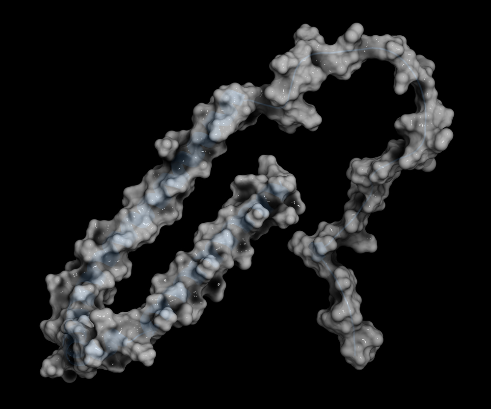
The difficulty in studying the protein aggregates is linked to their rapid formation and breakdown. When alpha-synuclein proteins misfold, they are prone to clump together with other misfolded proteins.
Scientists believe that these aggregates are most toxic when composed of a few proteins — what researchers call oligomers. But oligomers are unstable, and swiftly progress to form larger aggregates or break apart again.
To overcome this issue, researchers used advanced techniques, including a method called solid state nuclear magnetic resonance spectroscopy, to stabilize the oligomers long enough to study their structure and actions.
Looking at both toxic and non-toxic alpha synuclein oligomers in lab-grown rat neurons or human neurons gathered from cancer patients, the team noted how the oligomer binds to the cell surface and then breaks through the cell membrane to get attached to the wall of the neuron.
This triggers a series of events that eventually kills the cells.
“It is a common property of oligomers that they can damage brain cells once they bind to the surface,” said De Simone. “It is a bit like if you put a piece of extremely hot metal on to a plastic surface. In a fairly short space of time it will burn a hole through the plastic. The oligomer does something similar when it comes into contact with the cell membrane, and this disrupts the integrity of the membrane, which is the key step in the mechanisms leading to the death of the neuron.”
Their experiments also allowed them to identify the structural features of the oligomer that made this possible. Introducing mutations in the alpha-synuclein protein at the region that first binds to the cell membrane made the oligomers less toxic — an experiment that further confirmed their findings.
The information about key structures in the protein may allow researchers to develop drugs that target the interaction between alpha-synuclein and neuronal membranes.
“One of the really exciting things about this work is that not only has it been possible to define the structure of the critical pathogenic species in a neurodegenerative disorder, but we have also managed to propose a mechanism for its toxicity, providing vital clues for pursuing rational therapeutic strategies,” said Dobson, Master of St John’s College and a director of the Centre for Misfolding Diseases at the University of Cambridge.


Carlos
NOW THIS IS PROGRESS!!!!
Once we identify what is destroying Dopaminergiccells,WE'RE home!almost.
Prof. Pedro Moradas Ferreira
Although there is hope we know there is a long way to go before any compound preventing the formation of aggregates will be in the market
Thomas Leonard
Parkinson Patients: Take a look at taking Mannitol.