Parkinson-Related Research IDs Neurons That Control Movement
Written by |

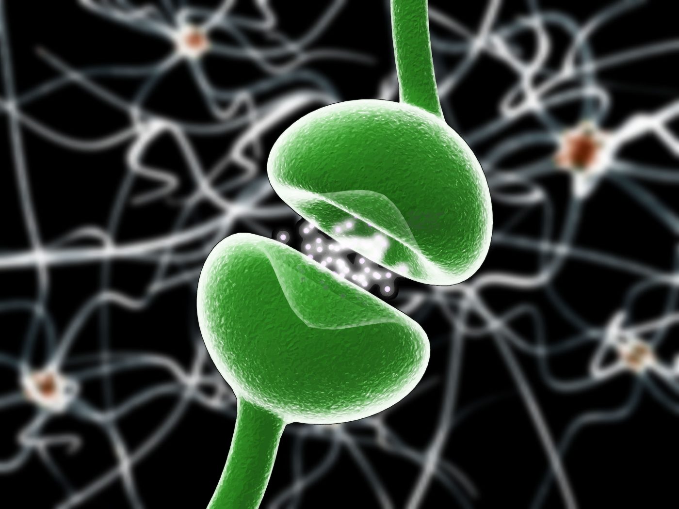
A team of researchers localized the specific types of neurons involved in movement, work that might be of relevance to understand neurodegenerative diseases like Parkinson’s and Huntington’s. The study, entitled “Cell-Type-Specific Sensorimotor Processing in Striatal Projection Neurons during Goal-Directed Behavior,” was published in Neuron.
Neurodegeneration occurs when brain nerve cells lose their functionality and, over time, ultimately die. In Parkinson’s and Huntington’s, for example, motor neurons of the central nervous system involved in movement and muscle coordination are damaged, causing symptoms like tremor, rigidity, slowness of movement, postural instability, and lack of coordination.
One key function of the brain is to analyze sensory information and make behavioral decisions (sensorimotor behavior) based on that information, like walking across a room or grasping an object. The brain region called the striatum, known as a critical component of the reward system, receives inputs from important neurotransmitters like glutamate and dopamine to serve as a coordination center of information analysis for various key cognitive activities, such as motor/action and decision making. Basically, the striatum contains various types of neurons, but little is known about their roles and functions, especially in movement.
In this study, the team examined mice striatum using cutting-edge technology. The animals were first trained to carry out simple goal-directed sensorimotor transformation — a person deflects a whisker, and the mice would lick a spout— so researchers could correlate behavioral performance with neuronal activity in the striatum by looking at changes in electrical activities occurring inside individual neurons. The results suggested that neurons extending from the striatum to a nearby part of the brain, named substantia nigra, are involved in movement. This small brain area has an important role in reward, addiction, and movement. To confirm the results, a reverse experiment was performed: Researchers activated neurons using optogenetics, a method employing a light-sensitive protein introduced into live neurons to stimulate and control the neurons. In fact, the data corroborated previous observations that stated neurons located in the brain regions that extend from striatum to substantia nigra were responsible for movement initiation. In other words, the researchers found that stimulating via optogenetics the same neurons in the striatum that affect the substantia nigra triggered the “lick spout” response without any external stimulous, like whisker deflecting.
These are significant results for neuroscience research, as they identify a specific type of neurons in the striatum where information turns into action, revealing a core function of the brain. In the long-term, this information could translate into possible treatments of neurodegenerative diseases, like Parkinson’s and Huntington’s, that affect action initiation and motor control.


