New Method Shows How Alpha-synuclein Can Damage Mitochondria
Written by |

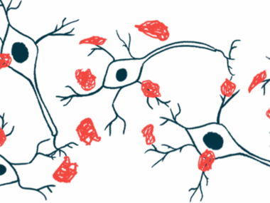
A new technique shows that alpha-synuclein, the protein at the heart of Parkinson’s disease, affects cell membranes differently, based on their composition.
This discovery sheds light on how alpha-synuclein clumps known as amyloids might disrupt cellular membranes, potentially helping to design therapies that might slow or stop the process and, by extension, disease progression.
Researchers at Chalmers University of Technology, in Sweden, adapted microscopy techniques used to analyze structural changes in biological molecules to understand how amyloid-forming proteins like alpha-synuclein can disrupt cellular membranes.
Their study, “Single-vesicle imaging reveals lipid-selective and stepwise membrane disruption by monomeric alpha-synuclein,” was published in the peer-reviewed journal PNAS.
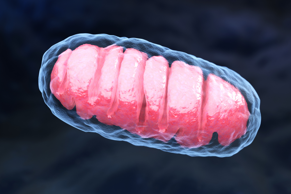
Mitochondria, membrane-enclosed structures within cells that produce energy, suffer damage over the course of Parkinson’s disease. Pathological versions of alpha-synuclein are thought to affect or even cause such damage.
To better understand this interaction, the Swedish team members investigated whether they could detect changes in membrane structure due to alpha-synuclein, which could lead to a more complete picture of the process.
The researchers generated two types of vesicles, or membrane-enclosed sacs, one of which approximated the composition of mitochondria and the other, that of synaptic vesicles, where alpha-synuclein is thought to help with release and trafficking.
Synaptic vesicles are tiny structures that store different neurotransmitters, or chemical messengers, that are then released at the synapse: the junction between two nerve cells that allows them to communicate.
The researchers then exposed these vesicles to small amounts of alpha-synuclein and monitored the resulting interactions.
Although the chemical differences in the two membranes were relatively small, alpha-synuclein had markedly different effects on each one. Alpha-synuclein attached itself to both membranes, but only deformed the mitochondrial-like membrane, causing its contents to leak out.
“In our study, we observed that alpha-synuclein binds to — and destroys — mitochondrial-like membranes, but there was no destruction of the membranes of synaptic-like vesicles,” Pernilla Wittung-Stafshede, PhD, professor of chemical biology at Chalmer University and one of the study’s authors, said in a university press release.
“The damage occurs at very low, nanomolar concentration, where the protein is only present as monomers — non-aggregated proteins,” she added. “Such low protein concentration has been hard to study before but the reactions we have detected now could be a crucial step in the course of the disease.”
An analysis of this interaction showed that to generate the observed membrane deformations, alpha-synuclein had to insert itself deeper into the mitochondria-like membrane than the synaptic vesicle-like membrane. Previously, alpha-synuclein was thought to lie flat upon the membrane surface.
This is consistent with observations that alpha-synuclein binds better to model membranes with low lipid density, high curvature, and/or those containing irregularities, such as is found in the mitochondria-like membrane.
Past studies have also reported that alpha-synuclein causes imperfections in these membranes by making them less rigid and more porous. The researchers mentioned that if alpha-synuclein binds preferentially to membranes that contain irregularities and can introduce irregularities on its own, then a small initial binding event could trigger a chain of events that leads to vesicle collapse.
This may explain why the team observed significant structural changes in response to a low number of alpha-synuclein molecules. They suggest that membrane regions with a relatively high density of alpha-synuclein molecules can make it easier for yet more molecules to attach themselves.
One of the key aspects of the team’s technique is that it enables researchers to study small numbers of molecules without the need for fluorescent markers. Fluorescent labeling is a common method for tracking specific biological molecules, but the labels themselves can affect the very reactions under study.
Having validated the utility of their technique, the researchers plan to experiment with alpha-synuclein variants and with membranes that more closely resemble those found in cells.
“We also want to perform quantitative analyses to understand, at a mechanistic level, how individual proteins gathering on the surface of the membrane can cause damage,” said Fredrik Höök, PhD, professor in the department of physics, who was also involved in the research.
“Our vision is to further refine the method so that we can study not only individual, small — 100 nanometres — lipid vesicles, but also track each protein one by one, even though they are only 1–2 nanometres in size,” he said. “That would help us reveal how small variations in properties of lipid membranes contribute to such a different response to protein binding as we now observed.”


