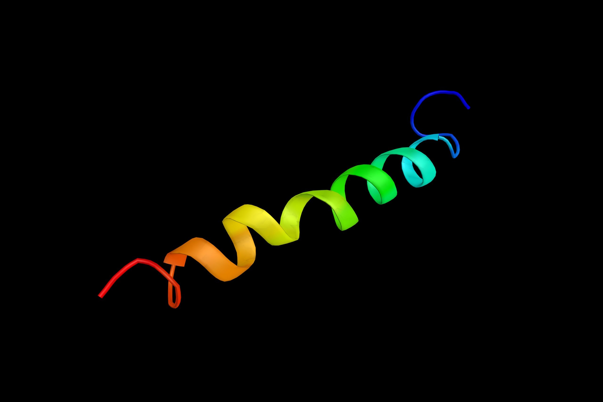
LRRK2 Protein’s 3D Structure Suggests New Way of Treating Parkinson’s
Written by |

A high-resolution 3D model of the leucine-rich repeat kinase 2 (LRRK2) protein provides some hints as to how this molecule participates in Parkinson’s disease — and suggests ways to sidestep its abnormal behavior, a new study reported.
In its active form, LRRK2 binds to structures of the cell’s cytoskeleton that are needed for molecular trafficking inside cells, arresting such molecular transport. Treatments that keep LRRK2 in its inactive form — and prevent this binding — are the most likely to show benefit in future clinical trials targeting this protein, the researchers noted.
Their study, “Structure of LRRK2 in Parkinson’s disease and model for microtubule interaction,” was published in Nature.
Parkinson’s, a progressive neurodegenerative disorder, results from the death of neurons that produce dopamine (dopaminergic neurons), a neurotransmitter needed to coordinate muscle movement. Loss of dopamine causes many of the disease’s symptoms, including tremors, loss of balance, and slowed movements.
Mutations in the LRRK2 gene are one of the most common genetic causes of Parkinson’s disease. The LRRK2 protein is also overly active in many Parkinson’s cases with no known genetic link.
This protein plays an important role in cellular metabolism, such as autophagy (the cell’s recycling and waste-disposal system), and the mitochondrial activity that provides energy to cells. Yet, researchers believe that the protein’s interaction with microtubules explains its role in Parkinson’s.
Microtubules are tiny filaments in cells that make part of their cytoskeleton. They also serve as a molecular highway system for transporting molecules within cells. Most Parkinson’s-causing LRRK2 mutations increase the amount of the LRRK2 protein that is bound to microtubules.
Researchers at the University of California San Diego attempted to understand how LRRK2 interacts with these structures, and how it affects the transport of cargo within cells.
The study was led by Samara Reck-Peterson, a PhD and cell biologist, and her collaborator Andres Leschziner. It focused on a specific region of LRRK2 — the kinase region — that the protein uses to activate its substrates (the substance it acts upon). Kinases control most processes that are disrupted in diseases, and researchers suspected this could also be happening with LRRK2 in Parkinson’s.
They found that LRRK2 essentially exists in two configurations, depending on whether it is active or inactive. “Think of Pac-Man with his mouth open,” Reck-Peterson said in a press release. In its active form, the kinase region is closed; in the inactive form, that region is open.
Notably, when the protein is closed, its 3D structure facilitates the formation of LRRK2 chains (made of many LRRK2 proteins bound to each other) that coil around microtubule filaments. The protein’s open, inactive form does not promote such protein chains.
The team then examined how these LRRK2 oligomers affected microtubule-based transport in cells. This was done by tracking the movement of two proteins — dynein and kinesin — that move on top of microtubules to transport cargo.
Results demonstrated that in its closed form, the kinase region of LRRK2 significantly inhibited dynein and kinesin movement, even at very low doses. The problem was not so much in their velocity, but rather in the length these proteins were able to move, suggesting that the LRRK2 chains surrounding the microtubules acted as roadblocks in microtubule transport.
While it’s not clear if these roadblocks contribute to neurodegenerative processes in Parkinson’s, the researchers tested which inhibitors of the LRRK2 kinase could best restore microtubule transport.
Currently, two classes of LRRK2 inhibitors are known. “Some drugs wedge into Pac-Man’s mouth, keeping it open, while others lock the mouth closed,” Reck-Peterson said.
Only the inhibitors that kept LRRK2 in an open configuration were found to restore dynein and kinesin movement. This is likely because the open LRRK2 proteins were not as able to form long LRRK2 filaments surrounding microtubules.
In contrast, inhibitors that arrested LRRK2 in a closed form increased its ability to form such filaments, and further diminish microtubule transport.
These findings are thought to offer important insights for clinical trials that target LRRK2 as a possible way of treating Parkinson’s.
“Our data have important implications for the design of LRRK2 kinase inhibitors for therapeutic purposes. They predict that inhibitors that favour the closed conformation of the kinase will promote LRRK2 filament formation, thus blocking microtubule-based trafficking, while inhibitors that favour an open conformation of the kinase will not,” the researchers wrote.
“These results should be taken into account to enhance therapeutically beneficial effects of LRRK2 kinase inhibition and to avoid potential unintended side effects,” they added.
LRRK2 inhibitors currently in trials work by keeping the kinase closed, and pharmaceutical companies might need to rethink their strategy, Reck-Peterson said.
“I think companies will be scratching their heads, thinking about developing inhibitors that leave LRRK2 off microtubules,” she added.


