
MRI in Parkinson’s Patients Revealed Elevated Levels of Iron in Different Brain Regions
Written by |
Researchers at Wenzhou Medical University in China recently reviewed literature related to iron levels in the brains of patients with Parkinson’s disease (PD) and confirmed elevated levels in several regions of the brain.
The review paper, “Meta-analysis of brain iron levels of Parkinson’s disease patients determined by postmortem and MRI measurements,” was published in Scientific Reports.
The deterioration and eventual death of motor neurons in Parkinson’s disease has been associated with many environmental and genetic causes, but the underlying mechanisms which cause the disease to develop are still unclear.
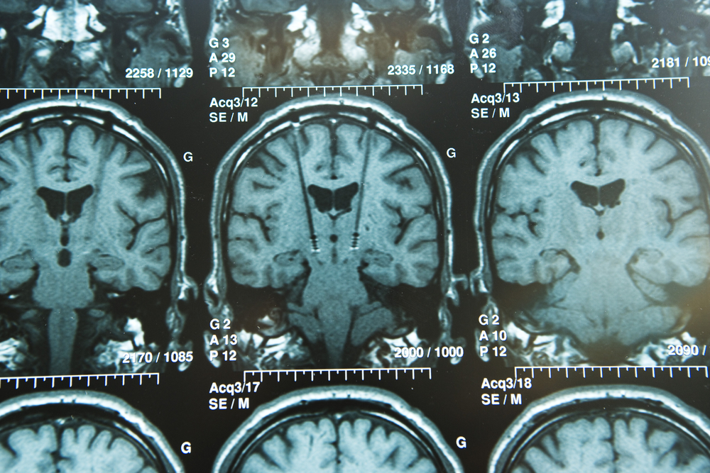
Several studies have linked iron overload to the disease course and development of Parkinson’s disease. Iron deposits have been observed in many brain regions of Parkinson’s patients using either postmortem spectroscopic analyses or magnetic resonance imaging (MRI) in real time.
R2/R2* relaxometry and susceptibility-weighted imaging (SWI) are two recent noninvasive techniques that can be used to estimate iron overload. However, despite the large number of studies, the methods are still not fully validated.
In this study, researchers reviewed available literature related to iron deposits in the brains of Parkinson’s patients in an effort to carry out a systematical meta-analysis.
They aimed to verify and confirm the presence of iron overload in brains of Parkinson’s patients; examine iron levels in different brain regions; and compare results of iron overload using two types of MRI (R2* relaxometry mapping and susceptibility-weighted imaging, or SWI), to postmortem samples.
They searched for articles published up Nov. 19, 2015, using different databases such as Medline, Web of Science, CENTRAL, and Embase. After examining thousands of articles, the researchers identified 33 that were relevant, of which 11 used postmortem analyses, 14 employed R2*, and 8 used SWI.
Analyses of the papers confirmed increased iron levels measured by postmortem spectroscopy, R2* or SWI imaging in the brain region of Parkinson’s patients called the substantia nigra, involved in reward and movement.
Both methods, postmortem and SWI, also showed significant iron accumulation in another brain region called the putamen, linked to regulation of movement and various types of learning.
The brain section called red nucleus, responsible for motor coordination, showed elevated deposits of iron using both R2* and SWI, while no data had been found in postmortem samples. Two other brain areas associated with motor processes, the nucleus caudatus and the globus pallidus, depicted significantly elevated levels of iron.
The researchers suggested that the presence of iron in these regions might indicate advanced disease progression, but the precise stage is difficult to determine.
“The current meta-analysis corroborates iron overload in substantia nigra and suggests such iron homeostasis defect in the putamen (by postmortem and SWI, but not R2*) and the red nucleus (by R2* and SWI; no data by postmortem) of PD patients,” the authors wrote.
“Both the R2* or SWI techniques may not authentically reflect iron changes in brain regions other than the substantia nigra,’’ they wrote.
“Our results offer a comprehensive understanding of iron loads in different brain regions in association with PD, and contribute to the evaluation of measuring accuracy of iron concentration by MRI methods,’’ they added.


Dusty Berry
I did not see anything where the PD patients levels of iron were compared to the levels in non-PD people in the same geographic area. Much more study needs to be done before claims are made.
Tim Bossie
It is true that a lot more study and research is needed for any kind of definite claims. However this information is from a study where researchers poured over other studies that have already been done where iron levels were compared.
DBParkinson's
Most research I have done indicates that excess accumulation of iron is more often related to dementia and cognitive dysfunction rather than pure movement disorder. Interestingly iron accumulation does not seem to correlate with ferritin and transferrin levels in the blood but more so with oxidative stress and the presence of Ferrous Sulfate. The presence of iron may actually differentiate between distinct sub-types of the disease rather than broad brushing it as an idiopathic feature. It may also in fact be a byproduct of the illness, not causative. More research is definitely required.
Tim Bossie
Thank you for adding your research to the mix. It is definitely quite interesting. But, you're right... more research is definitely needed into this.