MJFF funds collaboration to identify toxic proteins on PET scans
Companies developing imaging technology to help detect Parkinson's
Written by |
The Michael J. Fox Foundation (MJFF) is funding work to develop imaging technology for monitoring toxic proteins in Parkinson’s disease.
Xingimaging said it and its partner Synusight Biotech will use the $3.84 million grant to fund studies and work toward clinical application of their compound, 18F-FD4. The compound is a tracer molecule that targets toxic clumps of the protein alpha-synuclein — a characteristic sign and driver of Parkinson’s damage — designed to be used with positron emission tomography (PET) scans. This technique could help researchers and clinicians detect Parkinson’s earlier.
“We are deeply grateful for the support of the Michael J. Fox Foundation and share their commitment to developing a robust PET imaging biomarker targeting the core pathological hallmark of PD,” Roger Gunn, PhD, Xingimaging’s chief science officer, said in a company press release. “Such a biomarker would significantly enhance our understanding of the disease, its progression, and will play a central role in clinical trials evaluating new treatments.”
Parkinson’s disease is caused by the death of nerve cells that produce the signaling molecule dopamine. As these cells progressively die, various motor and nonmotor symptoms can develop. While several Parkinson’s treatments help manage the disease, there aren’t yet disease-modifying therapies that can slow or reverse progression.
Although researchers don’t exactly know why dopamine-producing cells die in Parkinson’s disease, toxic clumps of the alpha-synuclein protein are thought to play a role. As these clumps spread throughout the brain, they may disrupt nerve cell function. This process may begin before symptoms develop.

Identifying clumps may speed diagnosis, treatment
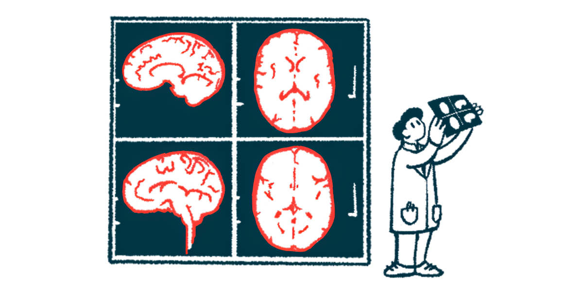
Identifying alpha-synuclein clumps in patients’ brains may help diagnose Parkinson’s early and help patients get treatment promptly, according to the companies. PET scans provide one possible route to monitor alpha-synuclein without requiring invasive procedures. The imaging method relies on radioactive tracing molecules that mark relevant proteins and processes.
Synusight developed the tracer FD4 to bind to alpha-synuclein aggregates. The company combined this with the radioactive molecule 18F, which allows the tracer to visually appear on PET scans. Together, they form 18F-FD4, “one of the most promising alpha-synuclein PET tracers in humans,” Gunn said.
In addition to supporting diagnosis, PET visualization of alpha-synuclein could help track disease progression and response to treatment.
18F-FD4 has shown signs of efficacy in preclinical and early clinical studies, according to Synusight. Investigator-initiated trials have tested the tracer in other diseases marked by alpha-synuclein clumps, including multiple system atrophy (MSA).
“Backed by the MJFF grant, we are now well-positioned to accelerate our validation efforts through additional clinical studies with XingImaging and fully unleash the potential of 18F-FD4 to advance the development of disease-modifying therapies,” said Roger Fan, CEO of Synusight. “Ultimately, we believe this tracer has the potential to transform the diagnosis and management of diseases and benefit millions of patients worldwide.”
Xingimaging is a contract research partner of Synusight and will support regulatory filings and clinical trials. The company develops PET imaging tools and provides brain imaging services.
Together, the collaborators aim to optimize methods for acquiring and analyzing 18F-FD4 imaging data. They will also use Xingimaging’s analysis pipelines to compare scans obtained from healthy people, Parkinson’s patients, and people with MSA.
“We continue to monitor the tremendous progress and advances in alpha-synuclein imaging,” said Jamie Eberling, PhD, senior vice president of research resources at MJFF. “XingImaging and SynuSight Biotech’s [18]F-FD4 programming is another hopeful step toward an urgently needed tool that could clearly measure, quantify and visualize brain pathology in Parkinson’s disease.”