Mouse Study Identifies Processes Responsible for Loss of Touch, Smell in Parkinson’s
Written by |

Researchers at the Karolinska Institute in Sweden have identified the underlying neural mechanisms responsible for the reduced sense of touch and smell which Parkinson’s patients experience in early stages of the disease. A better understanding of these processes can help in the development of new early diagnostic methods.
Their findings resulted from a study titled “Dopamine Depletion Impairs Bilateral Sensory Processing in the Striatum in a Pathway-Dependent Manner,” published in the scientific journal Neuron.
The primary recognized symptoms of Parkinson’s disease are muscular stiffness, poor balance, and uncontrollable trembling. Although there is no cure yet for Parkinson’s, many efforts have been made to reduce or delay these motor symptoms to improve patients’ quality of life.
In many cases the early signs of this disease are related not only to motor skills, but to sensory capacities, such as touch, smell, and vision impairments.
The motor symptoms of Parkinson’s have been related to the death of neurons and the loss of signals triggered by the neurotransmitter dopamine in a brain region called the striatum. But the cellular and neurological processes causing these sensory deficits were unclear to investigators.
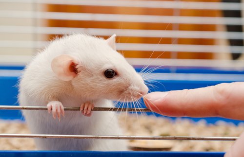
Making use of mouse models and a new tool called an optopatcher, researchers were able to record the striatum cells’ sensory response after they sent a light puff of air to either the right or left whiskers of mice. These sensory tests were performed in both normal mice and in mice lacking dopamine, which mimics the neurological networks found in Parkinson’s patients.
This approach allowed researchers to identify a specific neuronal network responsible for recognizing sensory signals in the striatum. This network was impaired in dopamine-deficient mice, which weren’t able to distinguish between left and right puffs of air to their whiskers. Those with normal dopamine levels presented an earlier and larger neurological response to the stimulation.
“By studying neuronal activity in the striatum, we found that the neurons in dopamine-depleted mice did not properly signal if it was the right or left whiskers that were being stimulated,” Gilad Silberberg, associate professor at Karolinska Institute’s Department of Neuroscience and senior author of the study, said in a press release.
The researchers were able to overcome the observed sensory deficits by treating the animals lacking dopamine with Levodopa (L-DOPA), one of the most commonly used drugs to treat Parkinson’s patients. The Levodopa treatments caused the mice to recover the ability to recognize left from right stimuli by restoring some of the features of the neuronal network.
This study not only highlights the involvement of this brain region in decoding sensory signals, it also described new processes involved in sensory impairments seen in Parkinson’s patients.


