
New Noninvasive Method Developed to Deliver Stem Cell Therapies for Neurological Diseases
Written by |

A new technology to deliver stem cell therapy to patients with neurological diseases such as Parkinson’s was developed by San Antonio scientists, according to a new study.
The study, “MRI Guided Delivery of Neural Stem Cells into the Basal Ganglia of Nonhuman Primates Reveals a Pulsatile Mode of Cell Dispersion,” was published in the journal Stem Cells Translational Medicine.
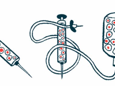
Stem cell therapy consists of the delivery of stem cells (immature cells that can become any type of cell in the body) to correct a deficiency in patients.
Marcel Daadi, associate scientist and director of the Regenerative Medicine and Aging Unit at Texas Biomedical Research Institute’s Southwest National Primate Research Center, and his colleagues had already developed stem cells capable of becoming dopaminergic neurons, which Parkinson’s patients lose over time.
“Stem cell-based therapy is emerging as a promising treatment for a variety of diseases and injuries,” the authors wrote. “The first step in evaluating the potential of different therapeutic stem cell lines is to develop a safe and effectively reproducible delivery system.”
Injection parameters have been studied in methods for drug delivery, but these cannot be directly applied to stem cell-based therapies. Also, the technology for stem cell delivery is still undeveloped and limited, and includes inconsistent cell survival, injection site inaccuracy, and the inability to avoid perforating structures like blood vessels, which could possibly cause a hemorrhage during stem cell delivery to the brain.
To avoid these limitations, the team tried to develop an MRI-guided technique as a potential method to implant stem cells into a living organism, hoping that one day this technique can also be applied to human patients.
The method was designed to deliver stem cells with low invasiveness and high accuracy in the brain region that controls motor skills, which are compromised in Parkinson’s patients, called the basal ganglia.
Testing the new technique in baboons, researchers observed that it not only provided effective targeted delivery, but also showed that, when injected in the brain, cells are not dispersed in a steady rate but instead in a small burst. This was a significant finding as it showed how injected cells disperse in the host brain and provide more information on how stem cell therapies can be designed to work best.
“We wouldn’t have been able to see this phenomenon using standard stereotaxic delivery,” Daadi said in a news release. “With iMRI, we can visualize in real time the cells being injected to the target area. A non-invasive iMRI approach is becoming a necessity in clinical applications to enhance the safety of patients and the efficacy of the therapeutic approach.”
“We can create the best cells, but if we can’t transplant them to the patient in a consistent and predictable way so that the patient can accept and thrive from them, then the therapy is simply ineffective,” he added.
The team believes this approach will improve the safety and effectiveness of stem cell transplants and address some of the current limitations in the delivery of stem cell therapies.

Michele Doyle
Wow!!This sounds very hopeful to those of us with Parkinsons. T iMRI procedure sounds like the DBS guided procedure that I had. Do you have any idea when this MIGHT be available??
Helen Ellevsen
I live in hope . I thrive on finding out what the scientific world is offering......lots of possibilities but how long is a piece of string. ......11 years with this diagnosis and I'm still leaving messages.
Is there no way possible for someone somewhere to end my nightmare.
Tim Bossie
We do hope that there is a day soon in which you can find relief. Stay strong and keep living in hope.
Trica
are any of these treatments available yet?