Impaired Movement in Parkinson’s Traced to Faulty Circuits in Brain Stem
Written by |

Researchers from the Gladstone Institutes published two research papers detailing what appears to be the mechanisms behind imbalances in specific brain regions that cause the movement and walking difficulties experienced by Parkinson’s patients. The papers appeared in the journals Cell and Neuron.
Faulty dopamine production is one of the hallmarks of Parkinson’s disease (PD) and has been the focus of a substantial portion of PD research. Depletion of this neurochemical occurs in the basal ganglia (BG), a region of the brain involved in processes such as movement and learning. In PD, there is an imbalance between two pathways in the BG, the “go” and “stop” pathways, which work together to control locomotion. Through mechanisms previously unknown, this balance fails in PD and the “stop” pathway overcomes the “go” pathway, making it difficult for patients to initiate a movement.
The first study, titled “Pathway-Specific Remodeling of Thalamostriatal Synapses in Parkinsonian Mice,” found that dopamine depletion causes a miscommunication between the BG and the thalamus, a region that is thought to forward sensory information between different subcortical areas and the cerebral cortex. This miscommunication leads to a loss of input in the “go” pathway from the thalamus, resulting in the loss of proper movement. By blocking the connection between the two regions, the scientists were able to reverse the imbalance between the two pathways and restore normal behavior in mouse models of PD.
“This study provides strong evidence for a mechanism by which the stop pathway overcomes the go pathway in Parkinson’s disease. Our findings implicate the thalamus in the development of the disease, an area of the brain that has received relatively little attention in Parkinson’s research,” Dr Philip Parker, the first author, said in a press release.
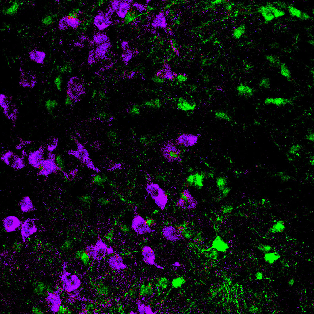
The second study, titled “Cell-Type-Specific Control of Brainstem Locomotor Circuits by Basal Ganglia,” uncovered the processes by which the “go” and “stop” mechanisms from the BG control locomotion. Researchers found these pathways regulate a group of nerve cells, located in the brain stem, that connects the brain to the spinal cord. Mice exercising on a treadmill were treated with a light tool that activated or inhibited certain brain cells, stimulating either the “go” or “stop” pathways. Results revealed the “go” pathway activated glutamate-producing neurons and that these cells were responsible for initiating locomotion. Activating the “stop” pathway inhibited these neurons, and movement in the animals effectively stopped. Moreover, signals from the brain stem could overpower those from the BG, meaning that even if the “stop” pathway is activated, mice would move if glutamate neurons were stimulated.
“In order to understand why walking is particularly disrupted in Parkinson’s disease, we need to map out the circuitry that controls locomotion,” said Dr. Anatol Kreitzer, the lead author of both studies. “Our study shows that a specific set of neurons in the brain stem are both necessary and sufficient to initiate locomotion. This finding could open the door for new treatment targets to help Parkinson’s patients walk more easily.”


