Fatigue Linked to Specific Patterns of Brain Degeneration in Parkinson’s Patients, Study Reports


In Parkinson’s disease patients, fatigue is linked to specific patterns of brain degeneration that are not present in individuals of the same age who do not have the condition, a study reports.
Findings of the study, “Structural brain correlates of fatigue in older adults with and without Parkinson’s disease,” were published in the journal NeuroImage: Clinical.
Previous research has shown that more than half of patients with Parkinson’s disease are affected by fatigue, which is one of the most common non-motor symptoms of the disorder and is also frequently found among older individuals without the disease. However, the reason why fatigue is so prevalent among older adults and whether it is linked to Parkinson’s is still unclear.
Because Parkinson’s disease is associated with structural changes in the brain, a group of researchers from the University of Colorado School of Medicine and collaborators set out to investigate if fatigue could be linked to alterations in the brains of Parkinson’s patients, compared with older individuals without the disease.
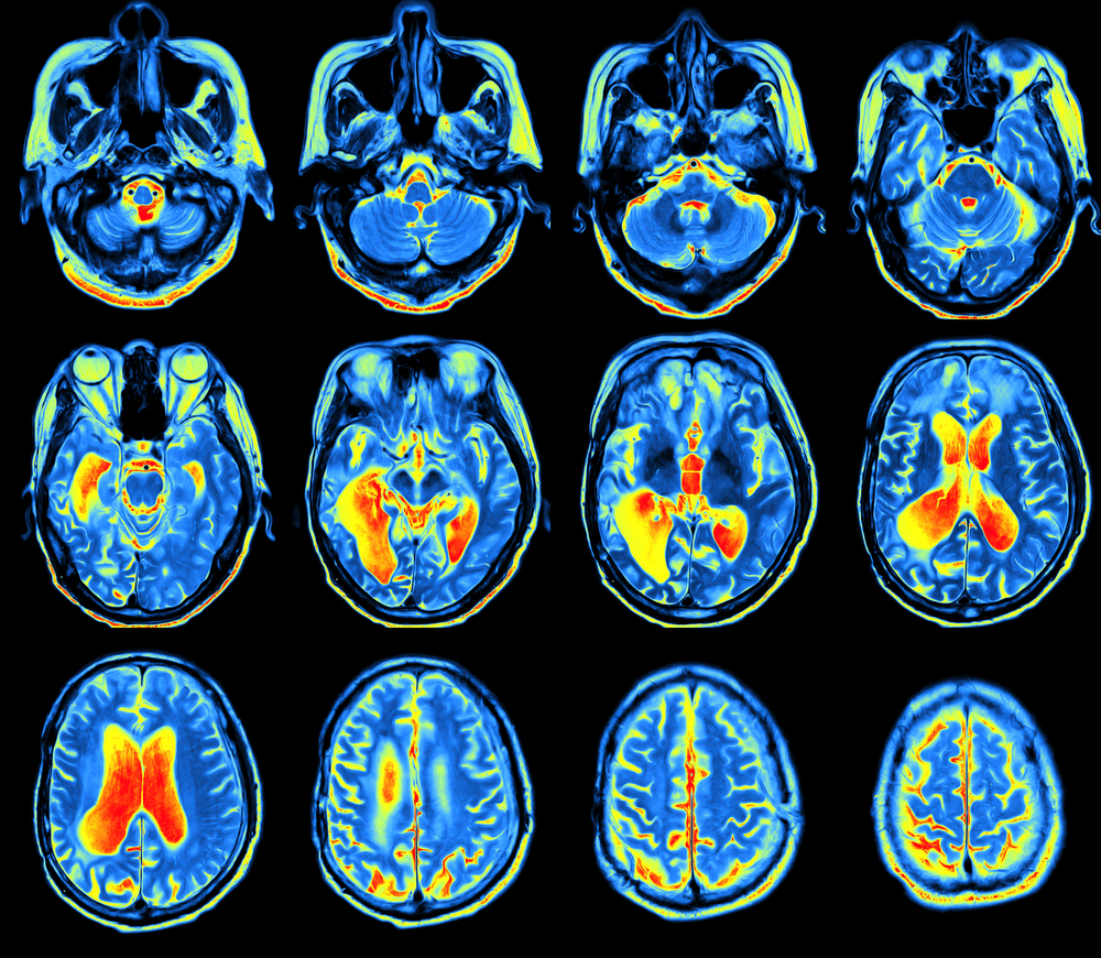
To study this, the team used a combination of magnetic resonance imaging (MRI) and diffusion tensor imaging (DTI) — a technique that measures the diffusion of water molecules in the brain to evaluate white matter integrity — to assess gray and white matter integrity.
The Parkinson’s Disease News Today forums are a place to connect with other patients, share tips and talk about the latest research. Check them out today!
Gray matter refers to areas of the central nervous system (brain, brainstem, and cerebellum) made up of neuron cell bodies, whereas white matter refers to areas of the central nervous system made up of myelinated nerve segments (axons) responsible for the transmission of nerve signals and that connect various gray matter areas.
The study involved a total of 60 patients with Parkinson’s — 17 women and 43 men with an average age of 67.58 years and mean disease duration of 5.67 years — and 41 age- and sex-matched healthy individuals used as controls.
Brain imaging data showed that Parkinson’s patients had a significant loss of gray matter volume in the brain’s striatum, a region involved in motor control, and the insula, a region involved in consciousness, emotion and motor control. The reduction in gray matter volume in the brain’s striatum was even more pronounced among patients who were also affected by fatigue.
No significant differences in brain structure were found between healthy controls with and without fatigue. Likewise, no significant differences were found on fractional anisotropy between those with or without fatigue from both groups. Fractional anisotropy reflects the diffusion of a liquid within white matter nerve tracts that connect gray matter areas.
“Fatigue in PD [Parkinson’s disease] is associated with unique [brain] structural changes, … suggesting fatigue in PD is primarily related to PD pathology [disease development], particularly in the dorsal striatum, and not simply a consequence of aging,” the researchers wrote.
“These results suggest that PD-related fatigue is a neurobiologically distinct syndrome and that further research is merited to better understand its phenomenology and pathophysiology with the goal of developing better treatments for this common and debilitating symptom,” they added.