New Scanner Worn as Helmet Allows Brain Imaging in Parkinson’s Patients


A new brain scanner that can be worn as a helmet could potentially revolutionize the world of human brain imaging, allowing patients with Parkinson’s disease to undergo brain scanning — a task previous traditional scanners failed.
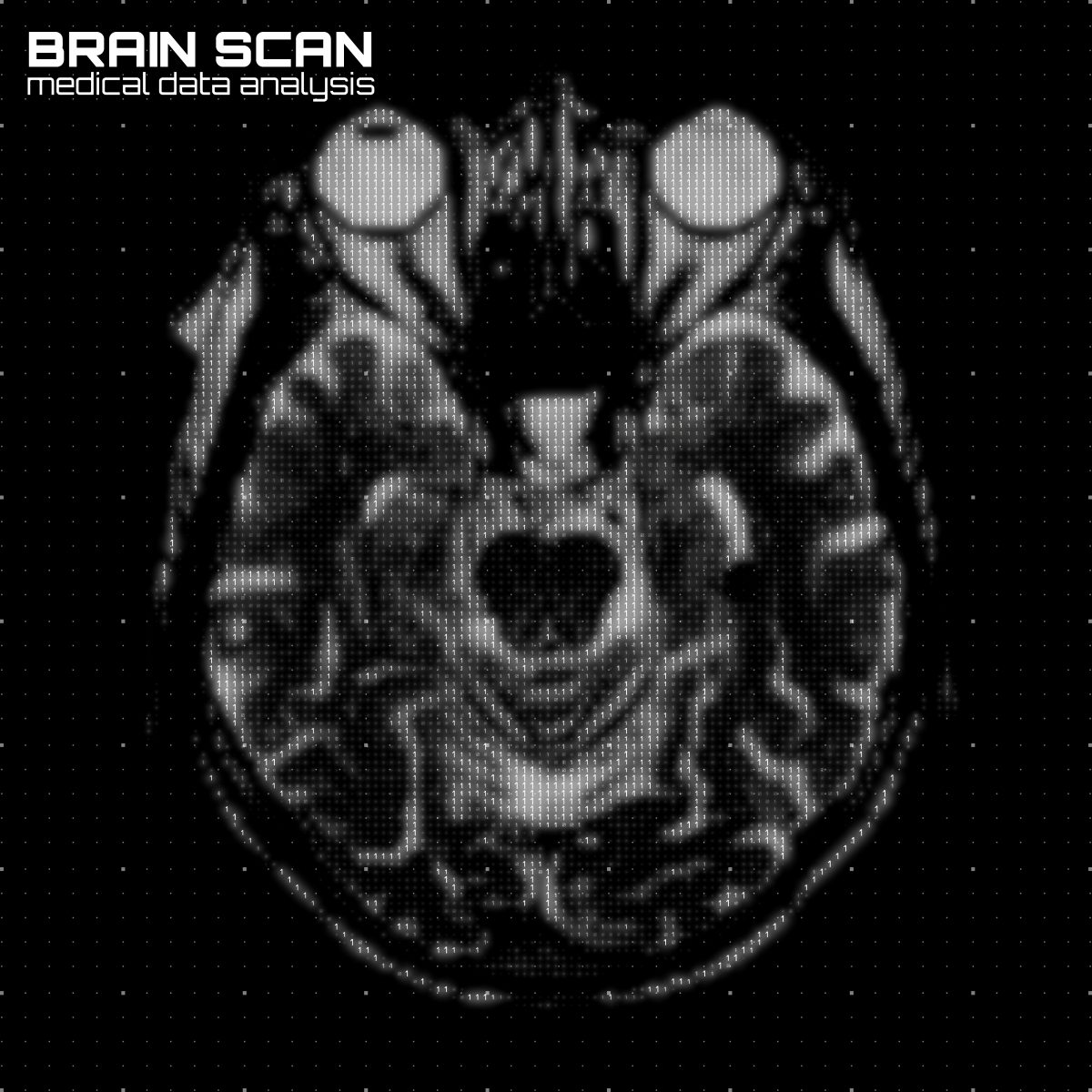
Brain cells use electrical impulses to communicate and, in doing so, form small magnetic fields that can be detected outside the head. Human brain function can be imaged by capturing these magnetic fields through a technique called magnetoencephalography (MEG).
With current scanners, patients have to remain perfectly still while being scanned, as even a tiny movement could render the images unusable. This means it is very difficult to scan people who find it hard to remain still, such as patients with movement disorders.
Researchers at the Sir Peter Mansfield Imaging Centre, University of Nottingham UK have tackled this issue by developing a new MEG system that’s worn as a helmet, allowing subjects to move around freely and naturally during scanning.
The new scanner is more sensitive than current systems, giving a detailed, millisecond-by-millisecond picture of the brain while subjects perform different tasks, such as speaking or moving.
It was recently described in the study, “Moving magnetoencephalography towards real-world applications with a wearable system,” published in the journal Nature.
“This new technology raises exciting new opportunities for a new generation of functional brain imaging. Being able to scan individuals whilst they move around offers new possibilities, for example to measure brain function during real world tasks, or genuine social interactions. This has significant potential for impact on our understanding of not only healthy brain function but also on a range of neurological, neurodegenerative and mental health conditions,” Matt Brookes, PhD, the study’s co-lead author, said in a press release.
Traditional scanners are extremely heavy, weighing about half a ton, because their sensors require extremely low temperatures (-269 degrees Celsius, equivalent to -452 degrees Fahrenheit), which means the machine must contain built-in cooling technology.
The new scanner has scaled-down technology that takes advantage of “quantum” sensors incorporated in a 3-D printed prototype helmet. The new sensors are extremely lightweight and can work at room temperature, allowing them to be placed directly onto the patient’s head. This positioning also means the sensors are closer to the brain, increasing the amount of signal they can pick up.
To allow patients to move their heads during scanning, researchers had to adjust the helmet’s electromagnetic potential and build special electromagnetic coils.
After designing a successful prototype, researchers are now developing new types of helmets to fit babies and children, as well as adults. They predict this new type of scanner will increase the sensitivity of brain imaging fourfold in adults, and up to 15 or 20 times in children.
“This has the potential to revolutionize the brain imaging field, and transform the scientific and clinical questions that can be addressed with human brain imaging. Our scanner can be worn on the head like a helmet, meaning people can undertake tasks whilst moving freely. Importantly, we will now be able to study brain function in many people who, up until now, have been extremely difficult to scan — including young children and patients with movement disorders. This will help us better understand healthy brain development in children, as well as the management of neurological and mental health disorders,” said Gareth Barnes, PhD, a professor and the leader of the project at Wellcome Centre for Human Neuroimaging at University College London.