Study Links Parkinson’s Patients’ Depression to Deterioration of White Matter in Their Brains


Researchers at China’s Nanjing Medical University have found a link between Parkinson’s patients’ depression and deterioration of white matter in their brains, particularly on the left side.
Their work, “Impaired long contact white matter fibers integrity is related to depression in Parkinson’s disease,” was published in the journal CNS Neuroscience and Therapeutics.
Brain tissue consists of gray and white matter. Gray matter includes synapses, or gaps between nerve cells that allows the cells to communicate with each others.
White matter is made up of nerve cell projections, known as axons or fibers, that connect distinct parts of gray matter. The length and condition of the fibers influences the way the brain processes information.
About half of Parkinson’s patients experience depression. Researchers have recently realized that it may not stem from despair over their disabilities but rather from the nerve cell degeneration their disease spawns.
White matter degeneration occurs frequently in Parkinson’s. The fiber connectivity problems stemming from the degradation can alter connections between brain regions involved in depression.
Using neuro-imaging, scientists have tried to identify white matter connections in the brains of depressed Parkinson’s patients. But most studies have focused on small brain areas, instead of the whole brain.
With that in mind, the Chinese team decided to study white matter fibers in depressed and non-depressed Parkinson’s patients.
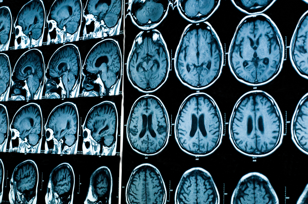
They did so with one of today’s most powerful magnetic resonance imaging (MRI) techniques: diffusion tensor imaging, or DTI.
DTI is a non-invasive way of understanding how brain structures are connected and fibers are organized. It is based on the rate of water molecule diffusion in the brain.
While molecules in a glass of water spread the same way in all directions, nervous system diffusion varies with the orientation of nerve cell fibers and the way they are organized — a phenomenon called fractional anisotropy.
Importantly, water diffusion patterns in brain regions change when the fibers are damaged.
Researchers analyzed water molecule diffusion patterns in 31 depressed and 37 non-depressed Parkinson’s patients.
They discovered a different diffusion pattern in regions on the left side of depressed patients’ brains than in the same regions of non-depressed patients’ brains. This was particularly true in long connection fibers in white matter.
Previous studies had suggested that the left side of the brain controls depression and anxiety, especially the front of the left side.
The same studies had found a connection between depression and abnormal diffusion patterns in two areas on the left side of the brain that previous studies had linked to depression — the left cingulum and left superior longitudinal fasciculus.
The Nanjing team called for studies of larger patient populations that use more comprehensive approaches “to reveal the unique features or imaging markers in depressed PD [Parkinson’s] patients. These findings may underlie the neural mechanisms of depression in PD and contribute to the diagnosis and treatment of depression in PD.”