New Ultrasound Technology for Treating Parkinson’s Disease Patients Under Clinical Trial
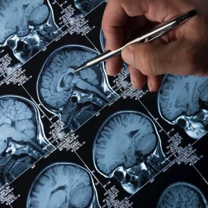


Researchers from the University of Maryland Medicine and from its Center for Metabolic Imaging and Image-Guided Therapeutics (CMIT) are conducting the first clinical trial with ultrasound waves to treat Parkinson’s disease (PD) patients. Using magnetic resonance imaging (MRI), they guide ultrasound waves through the intact skin and skull to a deep brain region, the globus pallidus. This structure regulates voluntary movement and can be targeted by medication and surgery to treat motor symptoms of tremor, rigidity and dyskinesia in patients with PD.
Levodopa is the current treatment for PD and can temporarily diminish motor symptomatology. However, in the long term, levodopa side-effects include involuntary movements called dyskinesias. Patients with advanced PD whose symptoms are not treatable by medication may undergo a surgical procedure known as deep brain stimulation. One of the brain regions stimulated by implanted electrodes is the globus pallidus though this surgery has associated risks.
Now, researchers have developed this new non-invasive treatment that guides ultrasounds to the targeted brain region by MRI. “In collaboration with my colleagues, we are excited to offer our patients a new, non-invasive therapy to control their Parkinson’s symptoms,” said principal investigator Howard M. Eisenberg in news release. “The neurology community has made significant strides in helping patients with Parkinson’s over the years; utilization of MRI-guided focused ultrasound could help limit the life-altering side effects like dyskinesia to make the disease more manageable and less debilitating,” added Dr. Eisenberg.
This new procedure lasts two to four hours with patients awake and lying on an MRI scanner with a head-immobilizing frame fitted with a transducer helmet. Ultrasonic energy is targeted through the skull to the globus pallidus of the brain, and images acquired in real-time. This allows the physicians to monitor the area being targeted and to make adjustments if necessary.
“We’re raising the temperature in a very restricted area of the brain to destroy tissue,” said Dr. Eisenberg. “The ultrasound waves create a heat lesion that we can monitor through MRI.”
Patients treated in the initial phase of the study experienced significant improvements in hand tremor.“Treatment-related side effects such as dyskinesia are the main reason my patients undergo surgery,” added Paul S- Fishman, sub-investigator on the clinical trial. “Focused ultrasound could offer these patients an alternative to surgery.”